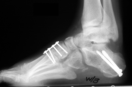

Imbalance between relatively weak peroneus brevis and preserved posterior tibialis muscle leads to inversion of calcaneus and adduction of foot. The extensor hallucis muscle which dorsiflexes the toe would then be unable to balance the combined plantarflexing forces of peroneus longus, posterior tibialis, and triceps surae muscles. This forefoot valgus position further drives the hindfoot into inversion. The relatively weak tibialis anterior is overpowered by the strong peroneus longus muscle which plantarflexes the first 1 st ray and pronates the forefoot (making forefoot valgus). Patho-anatomy and PhysiologyĬavus deformity of Charcot Marie Tooth disease is hypothesized to stem from asymmetric foot weakness anterior-laterally due to selective denervation of the tibialis anterior and peroneus brevis muscles of the legs. Severe pes cavus is 30% idiopathic in nature with 70% likely secondary to neurological causes, with most having origins with Charcot Marie Tooth disease (hereditary sensory motor neuropathy). Pes cavus occurs in about 8-15% of the general population.

Idiopathic subtle cavus feet without neurological disordersĮpidemiology and Risk factors for Prevention.Malunion of calcaneal or subtalar fracture.Posterior compartment syndrome of the leg.Hereditary sensory motor neuropathy (Charcot Marie Tooth disease) Regardless of the etiology of the neuropathy, intrinsic muscle atrophy and imbalance among different muscles group of the leg leads to features of pes cavus including high arch, clawing of the toes, and equinus deformity. Pes cavus cases can resultfrom hereditary or acquired peripheral neuropathies.

The presentations may merely be a result of abnormal biomechanics associated with cavus feet and identification and management of cavus feet is essential for successful treatment and avoidance of recurrence of symptoms. peroneal tendinitis) rather than deformity itself. Patients often present with painful conditions which result from pes cavus (e.g. Pes cavus may have concomitant hindfoot varus, equinus, forefoot adduction, forefoot valgus, and claw toe. Variations of pes cavus deformities exist and may be associated with acquired, hereditary, and congenital neurological or musculoskeletal conditions. Pes cavus is commonly characterized by its elevated longitudinal medial plantar arch of the foot and is also known as “claw foot, hollow foot, or cavovarus foot”.


 0 kommentar(er)
